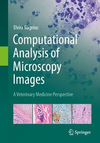
Computational Analysis of Microscopy Images
A Veterinary Medicine Perspective
- Publisher's listprice EUR 139.09
-
57 687 Ft (54 940 Ft + 5% VAT)
The price is estimated because at the time of ordering we do not know what conversion rates will apply to HUF / product currency when the book arrives. In case HUF is weaker, the price increases slightly, in case HUF is stronger, the price goes lower slightly.
- Discount 20% (cc. 11 537 Ft off)
- Discounted price 46 150 Ft (43 952 Ft + 5% VAT)
- Discount is valid until: 31 December 2025
Subcribe now and take benefit of a favourable price.
Subscribe
57 687 Ft

Availability
printed on demand
Why don't you give exact delivery time?
Delivery time is estimated on our previous experiences. We give estimations only, because we order from outside Hungary, and the delivery time mainly depends on how quickly the publisher supplies the book. Faster or slower deliveries both happen, but we do our best to supply as quickly as possible.
Product details:
- Publisher Springer Nature Switzerland
- Date of Publication 3 July 2025
- Number of Volumes 1 pieces, Book
- ISBN 9783031862694
- Binding Hardback
- No. of pages172 pages
- Size 240x168 mm
- Language English
- Illustrations XV, 172 p. 41 illus., 30 illus. in color. Illustrations, black & white 659
Categories
Long description:
"
This application-based guide fills a unique niche in the veterinary medical field by merging advanced computational techniques with the practical needs of veterinary pathology.
With increasing prevalence of digital pathology, there is a burgeoning requirement to navigate veterinary professionals in the utilization of computational methods and the enhancement of diagnostic accuracy. This book caters to this demand, presenting the material in an accessible way to novices, technologists, and pathologists. Written from the perspective of a seasoned veterinary pathologist, it ensures that the techniques described are relevant and directly usable.
Beginning with an exploration of microscopy fundamentals, the first part includes sample preparation, staining, and slide digitization. Subsequent chapters introduce readers to computational image analysis and the basics of image processing, tools, software, and successful integration of computational analysis into veterinary practice. Moreover, the book covers advanced topics such as image enhancement, reconstruction, quantitative analysis, and the application of machine learning and AI in microscopy image analysis. It provides insight into state-of-the-art imaging techniques like fluorescence and confocal microscopy, electron microscopy, and explores the innovations from nano to macro scales.
The incorporation of case studies and sample workflows allows this work to demonstrate the practical benefits of computational image analysis in veterinary medicine, with improvements in diagnostic accuracy and workflow efficiency. It serves as a learning resource for continuous professional development, helping veterinary pathologists stay abreast of technological advances in image analysis. Serving veterinary professionals, pathologists, researchers, and computational biologists alike, this book is an essential resource for anyone looking to harness the power of computational tools and AI in veterinary medicine.
" MoreTable of Contents:
"
Chapter 1. Computational Analysis in Veterinary Medicine.- Chapter 2. Fundamentals of Microscopy in Veterinary Pathology.- Chapter 3. Computational Image Analysis.- Chapter 4. Image Preprocessing Techniques.- Chapter 5. Image Segmentation and Feature Extraction.- Chapter 6. Quantitative Image Analysis in Veterinary Medicine.- Chapter 7. Machine Learning and AI in Microscopy Image Analysis.
" More


